
The motor cortex
*Motor areas of the cerebral cortex
1.The primary motor area (motor area 4 or MI)
2.The supplementary motor area (MII).
3.The premotor area (MIII).
4.The parietal lobe (somatosensory area).
1.The primary motor area:
Located in the pre-central gyrus.
Contain the highly excitable, large pyramidal cells, (Betz cells).
Control skeletal muscles of the opposite side of the body (Contralateral). EXCEPT for the upper part of the face, (forehead and the eyes) conrolled by both cerebral hemispheres).
Body representation is in an inverted manner (Upside down).
Cortical representation is proportional to the degree of skilled movement. Thus;
Areas representing muscles of speech & hands movement are large. while
Cortical area representing the trunk muscles is small.
Selective Lesion in (MI) lead to flaccid paralysis of the contralateral muscles,
due to loss of MI facilitation on Gamma Motor Neurons.
2- The supplementary motor area:
On the lateral side of the brain in front of area 4 and above the premotor area.
projects mainly to the motor cortex (planning and programming motor sequences).
3- The premotor area (MIII)
Representation in an inverted manner..
Stimulation of it→ complex coordinated movements, as setting the body in a certain posture to perform a specific task.
→ signals to the brain stem areas that regulate posture /or to M1 to excite multiple groups of muscles.
* It contains 4 specialized motor areas:
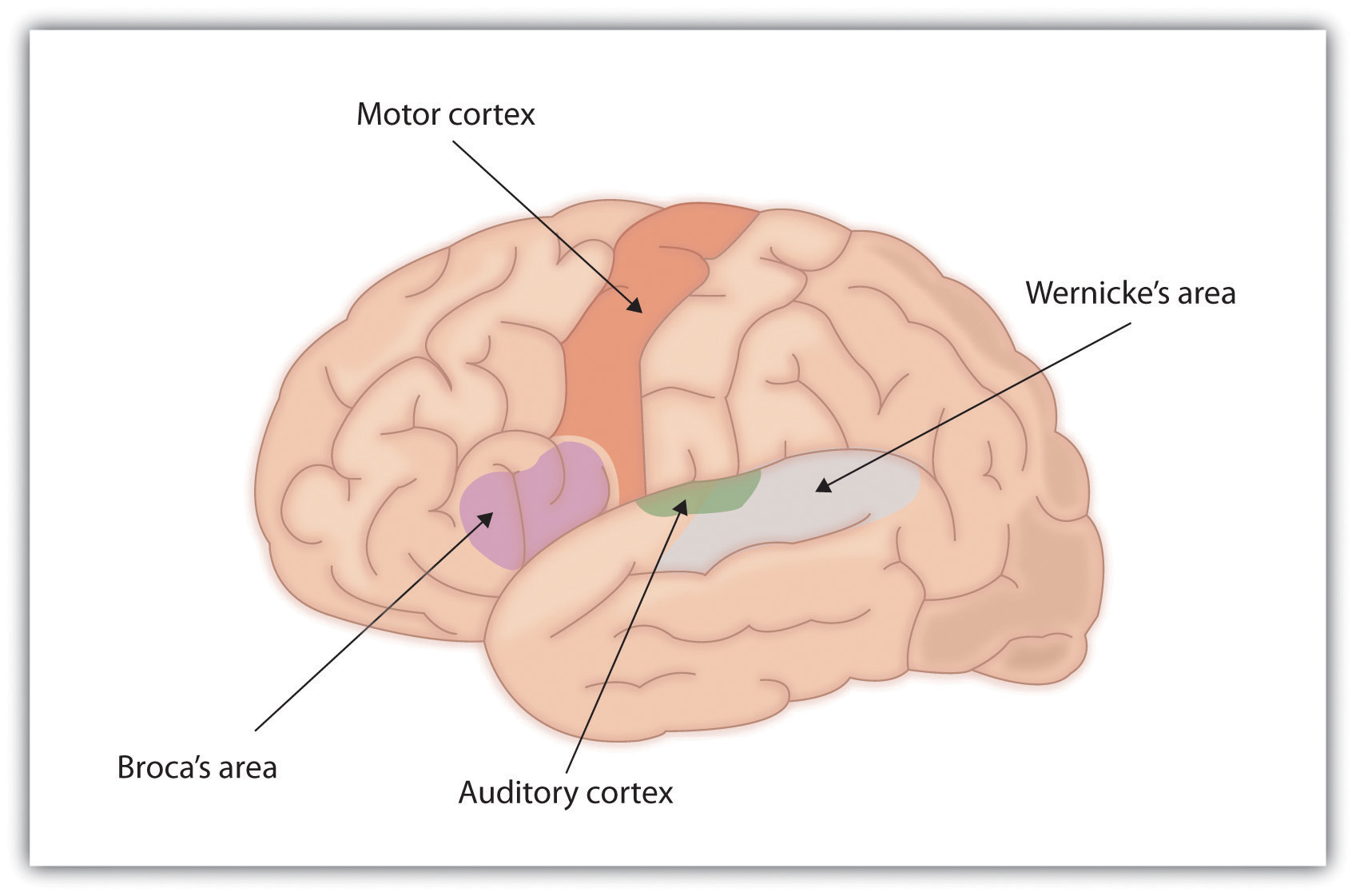
1. Broca's area (word formation centre):-
Immediately above the sylivian fissure.
Represent the tongue , lips and larynx
Its stimulation lead to vocalization.
Damage leads to motor (non-fluent) aphasia.
2. Voluntary eye movements area:
Lies above Broca's area.
Connected to occipital visual centers.
Control conjugate eye movements.
Damage prevents voluntary movement of the eyes towards different objects.
3. Head rotation area
Located above eye movements area, & connected with it,
Controls head rotation to wards objects.
4. The area for hand skills:-
Immediately anterior to the primary motor area M1 for the hands and fingers & above head rotation area.
*A lesion → motor apraxia.
Connections of the motor cortex:
1. Afferent fibers:-
>Fibers from near by cortical areas of the same hemisphere.
>Fibers from the corresponding motor areas of the other hemisphere via corpus callosum.
>Fibres from the ventroanterior & ventrolateral nuclei of the thalamus,these fibers relay impulses from the basal ganglia and cerebellum → co-ordination between cortex, basal ganglia and cerebellum.
>Fibres from the non-specific thalamic
nuclei.
* These fibers increase the general excitability of the cortex.
2. Efferent fibres:
>The descending tracts to the spinal cord
anterior horn cells and motor cranial nerve
nuclei ( i.e. the pyramidal and
extrapyramidal tracts).
> Also projects to the basal ganglia, the
cerebellum and brain stem.
*The pyramidal system
Include:
1. Corticospinal tract to the AHCs of spinal cord.
2. Corticonuclear tract to motor nuclei of cranial nerves.
> Consist of one millions fibres. As follow:
* 31% from primary motor area. Some
fibres are large myelinated, from the highly
excitable large pyramidal Betz cells of the
primary motor cortex Area 4.
* 29% from premotor area.
* 40% from somatic sensory areas 1.2.3.5,7 (posterior to the central sulcus).
> From the cortex the fibres descend into
the corona radiata → internal capsule
where corticonuclear fibers in the genu &
corticospinal fibers in the anterior 2/3 of the
posterior limb.
→ cerebral peduncle of the mid brain and
pons, to enter the medulla
> In the medulla , 80% of the corticospinal fibres cross to the opposite side
(pyramidal or motor decussation) & descend through the spinal cord as the crossed or lateral corticospinal tract.
>The fibers of the lateral corticospinal tracts supply the lateral neurons of the AHCs innervating the distal limb muscles of skilled movement.
> The remaining 20% of fibres descend to the spinal cord on the same side, as the direct or anterior (ventral) corticospinal tract. → Decussate to opposite side at the level of spinal cord segment .
* Interneurons that synapse with motor neurons in the medial part of the Anterior Horn Cell innervating the axial & proximal limb muscles of the posture control.
> These control the axial and proximal muscles of the limbs → posture.
> Ipsilateral fibers → bilateral innervation of some muscles on both sides of the body which move together (respiratory and abdominal muscles).
¤ The corticobulbar tracts:
> Made of pyramidal fibers that occupy the
genu of internal capsule & descend
through the brain stem→ cross to the
opposite side & synapse with motor nuclei
of cranial nerve supplying the muscles of the head.
Some of the fibers also synapse with
ipsilateral cranial motor nuclei → Bilateral
innervation of upper facial muscles.
> Functions of the pyramidal systems
Execution of complex, fine, skilled voluntary movements, especially of the fingers, toes and face.
> Facilitatory to stretch reflex & muscle tone .
Sectioning → Hypotonia.
> Some fibers pass directly to the
sensory relay nuclei of the dorsal horn
(corticofugal feed back pathways) That
modulates the intensities of incoming
sensory signals.
¤ The extrapyramidal system
>Made up of all parts in the CNS, that are
concerned with motor control, other than
the pyramidal system and cerebellum.
1- Cortical motor areas, especially the area 4s, premotor area and parietal cortex.
2- The basal ganglia.
3- Reticular formation, Red nuclei, Tectum of mid- brain and the Vestibular nuclei.
> Axons from the cortical areas descend in
the corona radiata and internal capsule,
intermingled (intermixed) with the
pyramidal fibres.
>In the basal ganglia, they synapse with
neurons of the caudata, putamen and
globus pallidus.
>The globus pallidus send impulses to the thalamus, red nucleus, substantia nigra, reticular formation, vestibular nucleus. tectum and inferior olive.
>From these areas, fibres descend to the
spinal cord, to supply the anterior horn
cells in the following tracts:-
1- Rubrospinal tracts: Is functionally associated with corticospinal system .
2- Reticulospinal tract.
3- Tectospinal tracts.
4- Vestibulospinal tracts.
5- Olivospinal tracts.
¤ Functions of the extrapyramidal system
Concern with control of :
>posture
>Planning , programming and initiation of movement,.
>Elicit the subconscious gross movements
associated with voluntary movements
, e.g. swinging of arms while walking.
*Some tracts are facilitatory to muscle tone (Vestibulospinal tract) while others are inhibitory (lateral reticulospinal tract).
*Have dominant inhibitory effect on gamma motor neurons to muscle spindle
Therefore damage lead to rigidity of muscles.


No comments:
Post a Comment